Brachyury Polyclonal / Unconjugated /
Brachyury Polyclonal / Unconjugated /
Product Details
| Supplier | R&D Systems | |
|---|---|---|
| Catalog #: | AF2085-SP (View supplier product page) | |
| Size | 25 μg | |
| Price | $169.00 | |
| Antigen | Brachyury | |
| Clone | ||
| Host | Goat | |
| Isotype | IgG | |
| Conjugate | Unconjugated | |
| Target Species | Chicken, Cynomolgus macaque, Human, Mouse | |
| Applications | WB, IHC, ChIP, FC, IHC-P, IHC-Fr, In Vivo, ICFC, ICC, ICC/IF | |
| Description | Human/Mouse Brachyury Antibody Intracellular Flow Cytometry = ICFC |
About Brachyury and Purified
| Brachyury | The protein encoded by this gene is an embryonic nuclear transcription factor that binds to a specific DNA element, the palindromic T-site. It binds through a region in its N-terminus, called the T-box, and effects transcription of genes required for mesoderm formation and differentiation. The protein is localized to notochord-derived cells. Variation in this gene was associated with susceptibility to neural tube defects and chordoma. A mutation in this gene was found in a family with sacral agenesis with vertebral anomalies. [provided by RefSeq, Sep 2018] |
|---|
Images

|
Detection of Brachyury-regulated Genes by Chromatin Immunoprecipitation. Mesoderm-differentiated BG01V human embryonic stem cells were fixed using formaldehyde, resuspended in lysis buffer, and sonicated to shear chromatin. Brachyury/DNA complexes were immunoprecipitated using 5 µg Goat Anti-Human/Mouse Brachyury Antigen Affinity-purified Polyclonal Antibody (Catalog # AF2085) or control antibody (Catalog # AB-108-C) for 15 minutes in an ultrasonic bath, followed by Biotinylated Anti-Goat IgG Secondary Antibody (Catalog # BAF109). Immunocomplexes were captured using 50 µL of MagCellect Streptavidin Ferrofluid (Catalog # MAG999) and DNA was purified using chelating resin solution. The VEGF promoter was detected by standard PCR. |
|

|
Brachyury in Differentiated Human Embryonic Stem Cells. Brachyury was detected in immersion fixed differentiated human embryonic stem cells using 10 µg/mL Goat Anti-Human Brachyury Antigen Affinity-purified Polyclonal Antibody (Catalog # AF2085) for 3 hours at room temperature. Cells were stained (green) and counter-stained with DAPI (blue). View our protocol for Fluorescent ICC Staining of Cells on Coverslips. |
|

|
Brachyury in Embryonic Mouse Notochord. Brachyury was detected in immersion fixed frozen sections of embryonic mouse notochord (E9.5) using 10 µg/mL Goat Anti-Human Brachyury Antigen Affinity-purified Polyclonal Antibody (Catalog # AF2085) overnight at 4 °C. Tissue was stained with the Northern-Lights™ 557-conjugated Anti-Goat IgG Secondary Antibody (red; Catalog # NL001) and counter-stained with DAPI (blue). View our protocol for Fluorescent IHC Staining of Frozen Tissue Sections. |
|
|
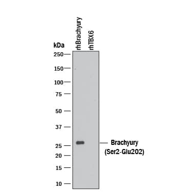
Detection of Recombinant Human Brachyury by Western Blot. Western blot shows 10 ng of Recombinant Human Brachyury and Recombinant Human TBX6. PVDF Membrane was probed with 0.1 µg/mL of Goat Anti-Human/Mouse Brachyury Antigen Affinity-purified Polyclonal Antibody (Catalog # AF2085) followed by HRP-conjugated Anti-Goat IgG Secondary Antibody (Catalog # HAF109). A specific band was detected for Brachyury at approximately 26 kDa (as indicated). This experiment was conducted under reducing conditions and using Immunoblot Buffer Group 3. |
|

|
Brachyury in BG01V Human Stem Cells. Brachyury was detected in immersion fixed BG01V human embryonic stem cells differentiated into mesoderm using Goat Anti-Human Brachyury Antigen Affinity-purified Polyclonal Antibody (Catalog # AF2085) at 10 µg/mL for 3 hours at room temperature. Cells were stained using the Northern-Lights™ 557-conjugated Anti-Goat IgG Secondary Antibody (red; Catalog # NL001) and counter-stained with DAPI (blue). Specific staining was localized to nuclei. View our protocol for Fluorescent ICC Staining of Cells on Coverslips. |
Citations
| Additional Sources |

|
|---|
Reviews & Ratings
Purified Excitation and Emission Spectra
Supplier Page